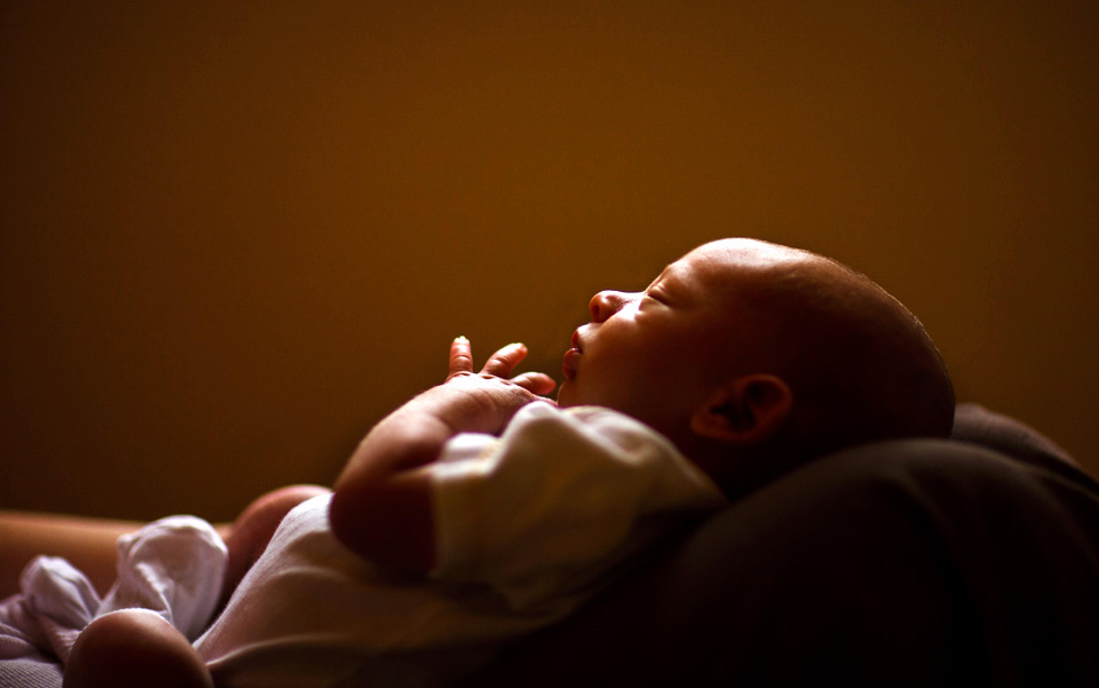
On a hot summer morning in Atlanta a few years ago, I took my then five-year-old son to his swimming lesson. As we walked toward his pool, we passed a smaller, shallower pool where an infant swim class was underway. I was still trying to wake up, but the class was already in full gear and my attention was drawn to a chorus of motherese (the high-pitched, rhythmic, infant-directed speech known more colloquially as baby talk) arising from the class. Parents were standing in a circle inside the pool, holding their infants in front of them, with the instructor in the centre. I couldn’t help but notice that many of the parents were fathers. Some were a bit chubby, with pale torsos reflecting the bright sunlight. They seemed like ideal infant-caregivers: calm, gentle, patient and sensitive. They didn’t seem like men you would go to battle with. In fact, they were the very antithesis of the warriors and athletes – think Maximus, Achilles or Michael Jordan – often associated with a masculine ideal.
Were the men in the pool just inherently different, born infant-caregivers? Or did the process of becoming a father somehow transform them so that they became better suited to perform this role? We have known for decades that mothers’ bodies and brains are transformed by the dramatic hormonal changes of pregnancy and childbirth. Now, new research is showing that men are also biologically transformed by the experience of becoming an involved father.
When women become mothers, levels of the hormones oestrogen, progesterone and prolactin increase throughout pregnancy. Hormones have their biological effects by binding to receptors – molecules that sense the hormonal signal – throughout the body, and they can influence behaviour through binding to receptors in the brain. Oestrogen increases the brain’s capacity to detect another major hormone, oxytocin, and the massive release of oxytocin at birth, coupled with repeated pulses of oxytocin during breastfeeding, helps mothers bond with and want to care for their infants.
But what about fathers? How do they get prepared to parent? This question was on my mind in 2008. I was teaching an undergraduate course on human social neuroscience. I included a unit on love, attachment and parenting, and was excited to use this class as an opportunity to update my knowledge in that area. In preparing for class, I found that there were many studies on the biology of human mothers, but I was struck by the dearth of studies at that time on the biology of human fathers. I knew that there was a decades-long trend of fathers becoming more involved as caregivers, but also that there was considerable variation in the extent of their involvement. I also knew that positive paternal engagement was associated with better social, psychological and educational outcomes in children, and I was aware of the tragic statistics of children raised without fathers. I decided then to shift the focus of my research, and my lab began investigating the biology of human fatherhood.
Our first study involved recruiting a large group of fathers with children one to three years of age. Our task was to compare their hormone levels and brain function with those of a group of men who were not fathers. We found that compared with non-fathers, fathers had 20 per cent less testosterone, the primary male sex hormone, often abbreviated as ‘T’.
The finding was fascinating in light of seminal research in birds, which had established that testosterone facilitated aggressive competition for mates and territory, while interfering with parenting. For example, when researchers gave monogamous, parental male birds extra testosterone, they became polygynous and non-parental.
This is an interesting change, but how strong is the evidence that a decrease in testosterone helps to shuttle energy from mating to parenting in species other than birds, including humans and other primates? Pretty significant, it turns out.
Marmoset monkeys are one of a minority of primate species in which adult males care for their offspring. Adult marmosets carry their twin infants on their back, a significant energetic burden. They also groom their infants and might share food with them. When the primate researcher Toni Ziegler and colleagues from the University of Wisconsin-Madison presented adult male marmosets with the scent of an ovulating female, testosterone levels skyrocketed, as if preparing the males for mating. However, this response was blunted in fathers compared with non-fathers, as if to discourage them from redirecting their attention from parenting. On the other hand, when Ziegler and colleagues presented adult males with the scent of an infant, testosterone levels decreased in fathers, as if preparing them to focus their energy on caregiving. By contrast, the infant scent had no effect on testosterone in non-fathers.
Such findings led to the idea that testosterone might redirect energy from parenting to mating in humans too. Important proof came in 2011, when the biological anthropologist Lee Gettler, then at Northwestern University, and colleagues measured testosterone levels twice in the same young Filipino men over a span of 4.5 years. Those who became fathers during that interval experienced a significantly larger decline in testosterone than men who didn’t become fathers, conclusively establishing that the transition to fatherhood decreases testosterone.
Skin-to-skin contact with premature infants increases both parental and infant oxytocin levels
The team also showed that Filipino fathers who were more involved in caregiving had a larger decrease in testosterone across the transition to fatherhood. And higher testosterone was associated with increased mating effort, as expected from the animal studies. For example, young men with higher testosterone levels at the initial measurement were more likely to find a mate and become fathers, a form of mating success, over the subsequent five years. What’s more, those fathers whose testosterone decreased the least across the transition to fatherhood reported the highest frequency of sexual intercourse and vice-versa.
Our own research into human fathers adds to the story, as well. We asked mothers to tell us how involved fathers were in caring for their toddler children, and found that men with higher testosterone tended to be less involved, whereas men with lower testosterone were more involved.
We made another important find. Despite having lower testosterone compared with non-fathers, fathers in our study actually had higher levels of another hormone that is classically identified with motherhood: oxytocin. In contrast with testosterone, oxytocin appears to promote paternal caregiving. In 2010, a Dutch research group led by the behavioural scientist Marian Bakermans-Kranenburg gave fathers oxytocin through a nasal spray and then watched them play with their child. When fathers were given oxytocin, they stimulated their child to explore more and showed less hostility toward the child compared with fathers who received a placebo treatment. Then in 2012, an Israeli group found that intranasal oxytocin made fathers touch their infants more and make more positive vocalisations towards them. In turn, infants, though not treated with oxytocin themselves, spent more time gazing at the father in this condition. Finally, and most remarkably, infants whose fathers were treated with oxytocin experienced an increase in oxytocin themselves. Thus, oxytocin seemed to initiate a positive feedback cycle that promoted father-infant bonding.
In 2016, another important study further implicated oxytocin in key aspects of paternal caregiving. Here, researchers measured the facial muscles of men watching infants express emotion. When the men were given oxytocin, their patterns better mimicked those of the infants, strongly suggesting that the hormone enhances paternal empathy.
In women, oxytocin is released by uterine contractions during labour and by nipple stimulation when breastfeeding. This begs the question, what triggers hormonal changes in men?
Researchers have found some answers here, too. There is evidence for a decline in fathers’ testosterone even during the partner’s pregnancy, so cues from the mother could be important. There is also evidence that postnatal contact with the infant can both lower T and increase oxytocin. Perhaps something about the appearance, the odour or actual tactile contact with the infant is responsible. A notable 2015 study showed that skin-to-skin contact with premature infants increases both parental and infant oxytocin levels. These findings predict that human fathers should become more strongly bonded to their children if they spend more time in close proximity to them as infants, and this has indeed been demonstrated.
To influence behaviour, hormones such as oxytocin must act in the brain. What is the brain’s actual neural circuitry that promotes paternal caregiving? Evidence points to a global parental caregiving system that generalises not only across mothers and fathers, but also across mammals. In the 1970s, the psychologist Michael Numan, then at the University of Chicago, discovered that damaging a particular small region at the base of the brain, known as the medial preoptic area or MPOA, severely disrupted maternal behaviour in laboratory rats. The MPOA is part of the limbic system, considered the ‘emotional’ brain, and resides within a structure known as the hypothalamus, a centre of sex and aggression, among other things.
Recently, a series of elegant experiments by the biologist Catherine Dulac and colleagues at Harvard University have investigated the role of the MPOA in the parental behaviour of male mice in great detail. Virgin male mice will attack and even kill pups, whereas mouse fathers are nurturing parents who lick and groom pups, build nests and retrieve pups to the nest.
Dulac and colleagues showed that both maternal and paternal behaviour activates a specific sub-population of neurons within the MPOA. Using sophisticated molecular techniques, they then selectively destroyed only this sub-population of MPOA neurons and found that it abolished both maternal and paternal behaviour and also elicited infanticide. On the other hand, when they activated these neurons in infanticidal males, it suppressed infanticide and elicited parental behaviour. Thus, the MPOA is a critical node in both the maternal and paternal brain.
Researchers have also shown that the MPOA activates a system of dopamine neurons projecting from the midbrain to a region known as the striatum, which is involved in reward and motivation. This motivational network is essential for parenting; damaging it inhibits caregiving, and rat mothers who lick and groom their pups more have more dopamine in the system.
In many species, this parental caregiving system is not active all the time. For example, female rats find pups unpleasant before giving birth, and require pregnancy hormones to activate the motivational system and stimulate maternal care. A prime player is oxytocin, released in the mother at birth and when nursing; it acts in both the MPOA and the midbrain to stimulate dopamine in the reward system, and this is presumably what makes pups rewarding and provides the motivation to deliver care. Rat mothers with more oxytocin receptors in the MPOA lick and groom their pups more; critically, their level of oxytocin receptors is influenced by the care that they received as a pup. Pups with more affectionate mothers have more oxytocin receptors. This might in fact be the mechanism for the transmission of parental caregiving styles from one generation to the next.
When fathers come into contact with their kids, it activates the midbrain, a region filled with dopamine neurons
Just as pregnancy hormones act on maternal brain circuits to stimulate caregiving, fatherhood can also alter the male brain so that parental caregiving circuits become more responsive to pups. The California mouse is another of the minority of mammalian species in which males are consistently involved in caring for the offspring. In 2003, the psychologist Brian Trainor then at the University of Wisconsin showed that an enzyme capable of converting testosterone into oestrogen increases in the MPOA when males become fathers. Critically, when they blocked the activity of that enzyme, they disrupted paternal behaviour, suggesting that MPOA oestrogen was essential for paternal behaviour. Other work has established that California mouse fathers also experience significant changes in the hippocampus, a brain region involved in learning and memory, and related structures. These include increased production of new neurons, changes in the shape of neurons that increase their ability to receive input from other neurons, increases in oestrogen receptors, and an increased number of oxytocin-containing neurons. Mandarin vole males also care for their offspring. When they become fathers, oxytocin receptors increase within the brain reward system, presumably rendering the system more sensitive to oxytocin so that pups become more rewarding.
My lab has been asking whether these animal models apply to human fathers as well. To find out, we adopted an ethological approach. Animal ethologists posit that offspring activate specific brain patterns and associated behaviours in parents. We sought to identify these patterns in human fathers by imaging their brains with functional magnetic resonance imaging (fMRI) while they responded to their children.
The early ethologists showed that young animals of all vertebrate species have a particular set of physical characteristics, called the baby schema, that tend to ‘release’ adult caregiving. These include large heads, protruding foreheads, large eyes, high brows, small lower faces and short, stubby limbs. We therefore reasoned that photographs of fathers’ children would function as releasers. Indeed, one important study showed that if you morph an infant’s picture to give it more baby schema (ie, you make the baby cuter), adults report stronger motivation to care for it and show stronger neural activation in the striatum. Another critical releaser of parental care that we used in our research is infant crying, which shapes parental behaviour through negative reinforcement. As my mentor Melvin Konner put it in his classic book, The Tangled Wing (2002): ‘It has to be a sound that sears its unpleasant way into the core of the mammal mother, causes a deep uneasiness and yet makes her deliver care instead of a lethal bite.’
As with other vertebrate parents, when human fathers come into contact with their offspring (in our experiment, through a photo) it activates the dopamine hub and the motivational system in the midbrain. The more the midbrain was activated, we found, the more involved the father was in caring for the child. This could mean that fathers who were more rewarded by their child became more involved in caregiving, or it could mean that, as fathers became more involved and formed stronger bonds with their child, they came to find the child more rewarding. Viewing pictures of their child also activated a number of other brain regions not included in animal models of parental brain function. These areas, including the anterior cingulate, the thalamus and the dorsomedial prefrontal cortex, all play a role in empathy. In humans, and likely many other species, parenting involves not only the motivation to deliver care but also the ability to perceive and understand the needs, feelings and mental states of the offspring.
We also found that supplemental oxytocin altered the way men’s brains responded to pictures of their children, boosting the response of the striatum, the target of midbrain dopamine neurons. It also increased activation in the anterior cingulate. Thus, oxytocin could render the child a more rewarding stimulus and might also increase paternal empathy.
In more recent research, we studied the effect of infant crying, that aversive stimulus that effectively forces the caregiver to share in an offspring’s pain or figure out a way to alleviate it. Unsurprisingly, infant crying strongly activates brain regions involved in empathy, such as the anterior insula and the anterior cingulate cortex, especially in more empathic fathers.
Paradoxically, we found that fathers who activate the anterior cingulate the most when listening to infant crying report the most negative emotional responses to those cries, in particular being more likely to label the cry as spoiled or manipulative. How can greater engagement of a brain region associated with empathy be linked with such negative subjective reactions to the cry? We suspect it relates to a phenomenon known as ‘empathic overarousal’, in which an observer takes on the distress of another individual to such an extent that they become mired in personal distress, which in turn interferes with the motivation and ability to deliver compassionate care. There might be an optimal state of arousal and degree of empathy, neither too high nor too low, that facilitates sensitive and responsive fathering.
Our group also evaluated the hypothesis that the trade-off between male mating and parenting occurs at the level of the brain. To do so, we compared the neural response of fathers and non-fathers for both child and visual sexual stimuli. We found that fathers had a stronger neural response to pictures of children than did non-fathers in brain regions involved in reward processing, suggesting that they might attach greater value to those stimuli. On the other hand, and consistent with the hypothesis, fathers had a weaker response to visual sexual stimuli than did non-fathers in brain reward regions.
Our prefrontal cortex allows us to override ancient impulses in the service of honouring commitments
Since male testes make sperm, testis size can be yet another measure of investment in mating effort. In primate species with promiscuous mating systems, in which females mate with multiple males and vice versa, males tend to have large testes for their body size. This is presumably so that males can produce large numbers of sperm cells that can compete with the sperm of other males inside the female reproductive tract to fertilise the egg. On the other hand, in species where females mate with just one male, males have small testes for their body size.
In light of this, we used MRI scans to measure testes size in human males, and asked if it was related to either parental behaviour or brain function. We found that males with smaller testes were both more involved in caregiving and had a stronger response to viewing pictures of their child within midbrain areas involved in parental motivation. While statistically significant, this correlation between testis size and caregiving was weak, implying that most of the variation in caregiving was explained by other factors such as occupational demands, availability of other caregivers, and social and cultural expectations. Nevertheless, these results provide further support for the notion of a trade-off between mating and parenting effort.
Our research suggests similarity between the hormonal and neurobiological underpinnings of caregiving in mothers and fathers. But there are differences as well. As one example, the neuropeptide vasopressin, a close molecular analogue of oxytocin, could be particularly important for paternal caregiving. For example, injecting vasopressin into the brains of virgin male prairie voles elicits paternal behaviour that involves contacting and crouching over pups, whereas blocking vasopressin action at its receptor decreased licking and grooming of pups. In marmoset monkeys, fathers have more vasopressin receptors in their cerebral cortex compared with marmoset males who are not fathers. There has been only limited work on the role of vasopressin in human paternal behaviour, but one study showed that giving vasopressin increased the amount of time that expecting fathers spent watching baby-related avatars in a virtual reality environment.
Finally, there is one crucial paternal brain region that we have so far neglected. Evolutionary theory predicts that males will invest more energy in mating versus parenting if that is how they can maximise their reproductive success, and it is natural for us to ask if the theory applies to our own species. Some men behave much as theory would predict. Consider a man who leaves his menopausal wife and family to start a new family with a younger, fertile woman. Or think of certain high-status, married fathers who spend considerable time and money on girlfriends, mistresses and even prostitutes. Yet, many other men choose to forego these pursuits. They override impulses that evolution has programmed into their brains, impulses that evolved because they enhanced the reproductive success of their ancestors. They do so out of love and respect for their partners and their children, and out of respect for social and cultural norms. But how do they do what males of other species seem incapable of?
The answer, I believe, is that they rely on the crowning achievement of human brain evolution: the prefrontal cortex. Not only is the human brain three times larger than the brain of our closest living primate relatives, the great apes, but the human prefrontal cortex is also larger than expected for our brain size. Our prefrontal cortex is what allows us to override ancient, evolved impulses in the service of honouring commitments, abiding by social norms, and exercising our moral responsibilities. We are privileged to have this remarkable organ, and we fathers would all do well to make use of it.
