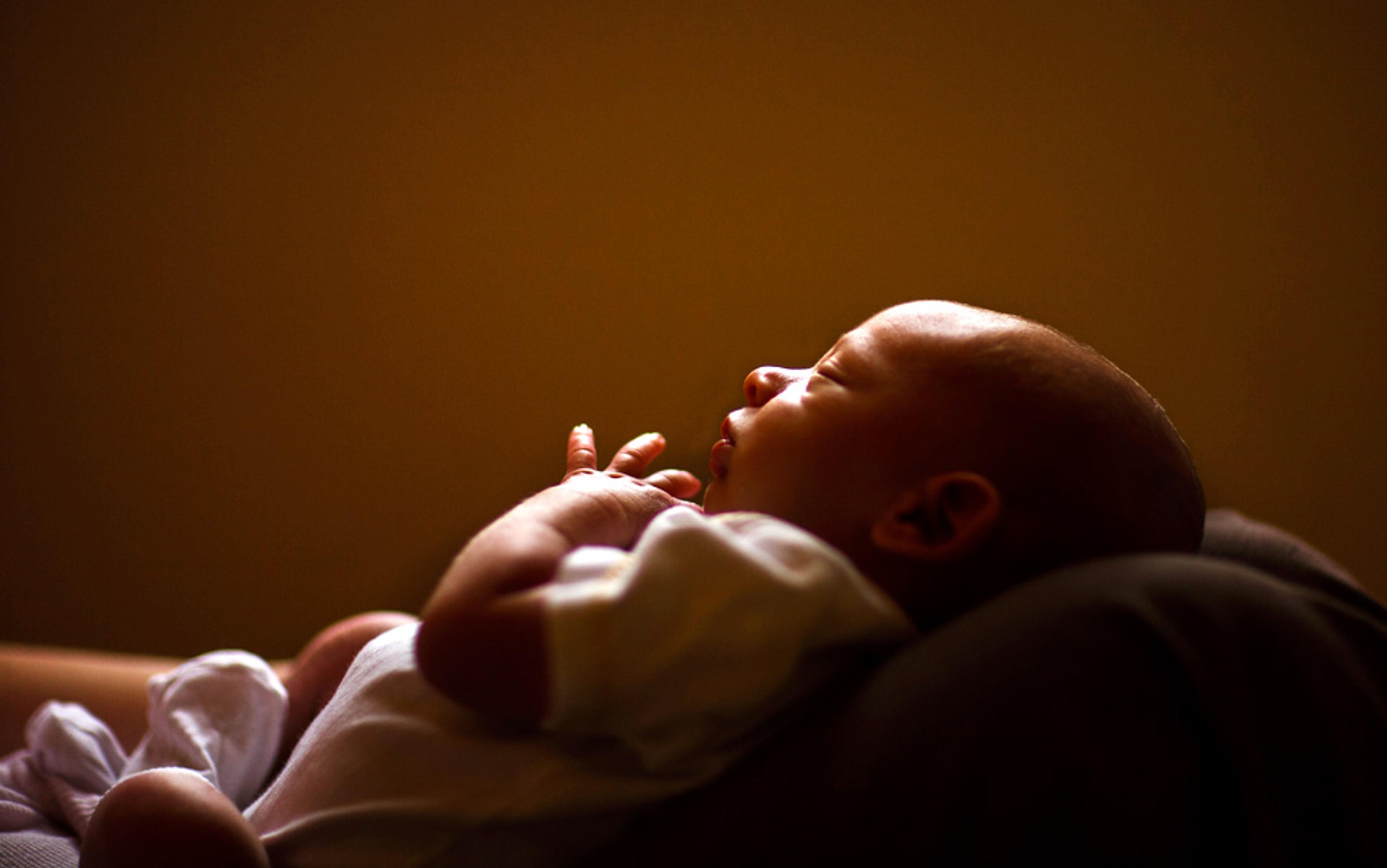
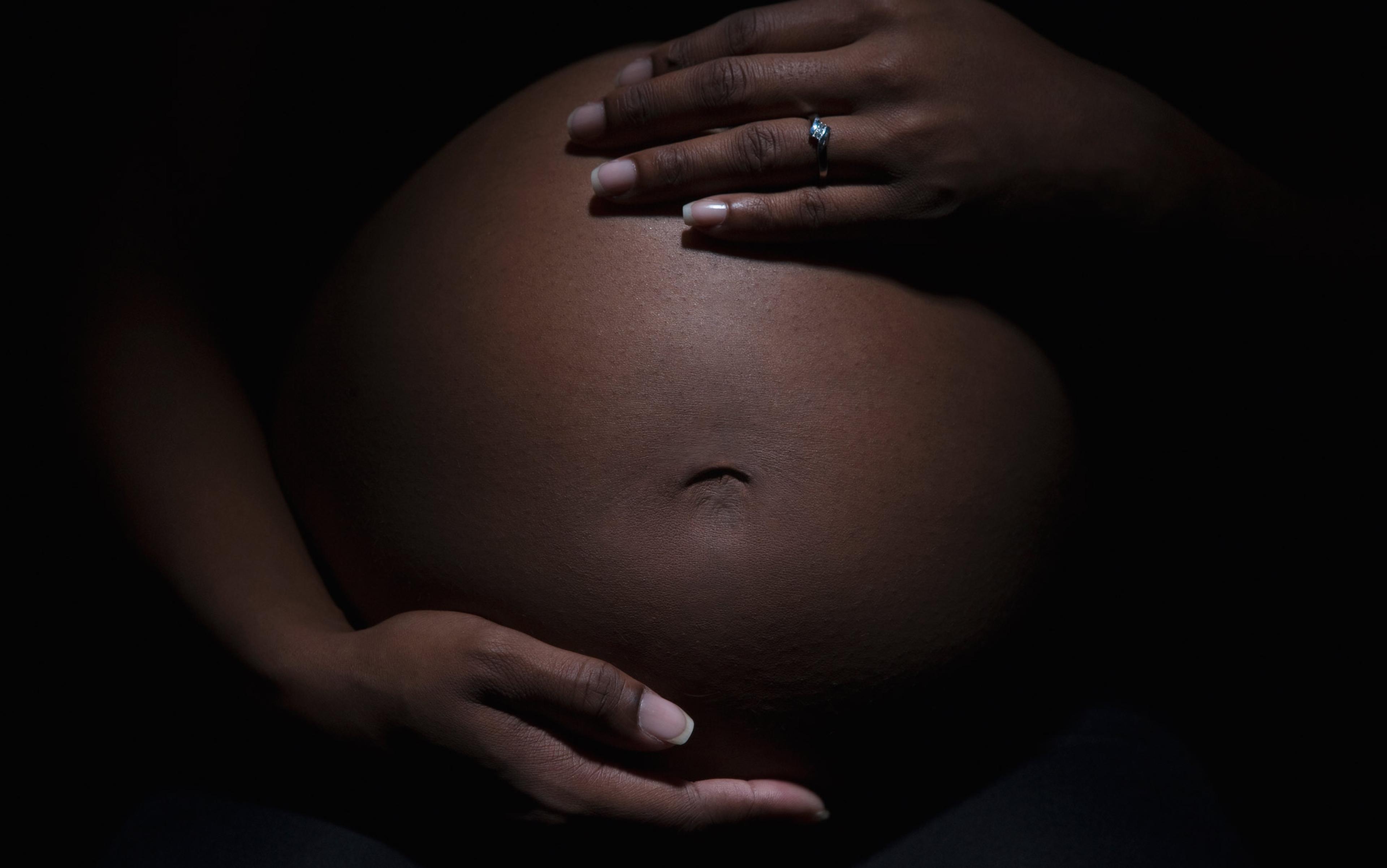
When Lee Nelson first began researching autoimmune disorders in the 1980s, the prevailing assumption was that conditions such as arthritis and lupus tend to show up more commonly in women because they are linked to female sex hormones. But to Nelson, a rheumatologist at the Fred Hutchinson Cancer Research Center in Seattle, this explanation did not make sense. If hormones were the culprit, one would expect these afflictions to peak during a woman’s prime reproductive years, when instead they typically appear later in life.
One day in 1994, a colleague specialising in prenatal diagnosis called her up to say that a blood sample from a female technician in his lab was found to contain male DNA a full year after the birth of her son. ‘It set off a light bulb,’ Nelson told me. ‘I wondered what the consequences might be of harbouring these lingering cells.’ Since the developing foetus is genetically half-foreign to the mother, Nelson set out to investigate whether it could be that pregnancy poses a long-term challenge to women’s health.
Evidence that cells travel from the developing foetus into the mother dates back to 1893, when the German pathologist Georg Schmorl found signs of these genetic remnants in women who had died of pregnancy-induced hypertensive disorder. Autopsies revealed ‘giant’ and ‘very particular’ cells in the lungs, which he theorised had been transported as foreign bodies, originating in the placenta. While Schmorl speculated that this sort of cellular transfer also took place during healthy pregnancies, it was not until more than a century later that researchers realised that these migrant cells, crossing from the foetus to the mother, could survive indefinitely.
Within weeks of conception, cells from both mother and foetus traffic back and forth across the placenta, resulting in one becoming a part of the other. During pregnancy, as much as 10 per cent of the free-floating DNA in the mother’s bloodstream comes from the foetus, and while these numbers drop precipitously after birth, some cells remain. Children, in turn, carry a population of cells acquired from their mothers that can persist well into adulthood, and in the case of females might inform the health of their own offspring. And the foetus need not come to full term to leave its lasting imprint on the mother: a woman who had a miscarriage or terminated a pregnancy will still harbour foetal cells. With each successive conception, the mother’s reservoir of foreign material grows deeper and more complex, with further opportunities to transfer cells from older siblings to younger children, or even across multiple generations.
Far from drifting at random, human and animal studies have found foetal origin cells in the mother’s bloodstream, skin and all major organs, even showing up as part of the beating heart. This passage means that women carry at least three unique cell populations in their bodies – their own, their mother’s, and their child’s – creating what biologists term a microchimera, named for the Greek fire-breathing monster with the head of a lion, the body of a goat, and the tail of a serpent.
Microchimerism is not unique to pregnancy. Researchers realised in the 1990s that it also occurs during organ transplantation, where the genetic match between donor and recipient determines whether the body accepts or rejects the grafted tissue, or if it triggers disease. The body’s default tendency to reject foreign material begs the question of how, and why, microchimeric cells picked up during pregnancy linger on indefinitely. No one fully understands why these ‘interlopers’, as Nelson calls them, are tolerated for decades. One explanation is that they are stem or stem-like cells that are absorbed into the different features of the body’s internal landscape, able to bypass immune defences because they are half-identical to the mother’s own cell population. Another is that pregnancy itself changes the immune identity of the mother, altering the composition of what some researchers have dubbed the ‘microchiome’, making her more tolerant of foreign cells.
The phenomenon, believed to have developed in mammals some 93 million years ago, is common to placental mammals to this day. Its persistence and reach was made astonishingly clear in 2012, when Nelson and colleagues analysed brain samples drawn from dozens of deceased women, ranging in age from 32 to 101. They found that the majority contained male DNA, presumably picked up from past pregnancies. And some of these Y chromosome cells had apparently been there for decades: the oldest subject was 94, meaning that male DNA that transferred during gestation would have persisted for more than half a century.
Most of the research focuses on the Y chromosome as a marker for foetal microchimerism. This does not mean that sons, rather than daughters, uniquely affect their mother’s bodies, but rather reflects an ease of measurement: the Y chromosome stands out among a woman’s XX genes. And there is nothing to suggest that the presence of male cells in women’s brains wields a particular influence. Nonetheless, the findings gesture toward an array of questions about what it means for one individual to play host to the cellular material of another, prompting scientists to look into whether this phenomenon affects physical health or influences behaviour, or even carries metaphysical consequences. The Western self is a bounded, autonomous entity, defined in no small part by its presumed distinction from the other. But this unfolding field of research, advanced by Nelson and others, suggests that we humans are not oppositional but constituent beings, made of many. Nelson, who is fond of referencing the poet Walt Whitman’s multitudes, says we need a ‘new paradigm of the biological self’.
One of the most cherished images in the West – if not in the world – is the mother and child. Gazes intermingled, bonded as if one, they are suspended in serene togetherness. This cooing placidity presents a scene of utter naturalness, of womanhood fulfilled, of tender destiny. In 1884, the physician John Harvey Kellogg urged women – at a time when childbirth was a leading cause of women’s death – to opt for the ‘slight inconveniences of normal pregnancy and physiological childbirth rather than the dismal comfort of a childless old age’. In spite of the acute health risks that gestation and delivery entailed for Western women well into the 20th century, pregnancy was commonly depicted as the ultimate form of cooperation – mothers sharing their bodies to the point of sacrifice for the sake of kin and species.
This vision utterly obscures the fraught evolutionary journey that delivers the babe in arms, and the screaming, nerve-jangled moments that surround it. Increasingly, pregnancy has come under scrutiny for its profound paradoxes. It is at once essential and unrivalled in its perils. As it engenders life, it also results in staggeringly high rates of death and disease. Scientists are starting to look to microchimerism for clues as to why pregnancy is both life-giving and a singular source of risk.
On one side of the spectrum, foetal microchimeric cells have been implicated in autoimmune disorders, certain cancers and pre-eclampsia, a potentially fatal condition characterised by high blood-pressure during the latter half of pregnancy. But another body of research has found that foetal cells can protect the mother. They appear to congregate at wound sites, including Caesarean incisions, to speed up healing. They participate in angiogenesis, the creation of new blood vessels. A recent survey of the immunological implications of microchimerism in Nature Reviews by researchers at the Cincinnati Children’s Hospital asserts that these cells ‘are not accidental “souvenirs” of pregnancy, but are purposefully retained within mothers and their offspring to promote genetic fitness by improving the outcome of future pregnancies’. The researchers suggest that microchimeric cells boost the mother’s tolerance of successive pregnancies, representing an ‘altruistic act of first children’ to support the success of their genetically similar siblings. And they are associated with decreased risk of Alzheimer’s, lower risk of some cancer, and improved immune surveillance – that is, the body’s ability to recognise and stave off pathogens. According to Nelson, having a different set of genes provides ‘a different looking glass for detecting a pre-malignant cell’.
Although foetal cells might contribute to certain autoimmune disorders, they could also benefit women with rheumatoid arthritis. While doctors have been aware since the early 20th century that arthritic pain tends to recede with pregnancy, Nelson and her colleagues wondered whether there is an immunological reason why it tends to re-emerge later. They found that higher levels of microchimerism were associated with a lessening of symptoms, and that giving birth offered a long-term protective benefit. ‘It really looks vaccine-like,’ Nelson said, noting that pregnancy provides temporary protection against rheumatoid arthritis that, much like a vaccine, diminishes over time. ‘Protection starts about a year after birth, and then gradually attenuates after about 15 years.’
‘There is definitely an association between the presence of foetal cells and improved disease status’
Foetal microchimeric cells might even extend longevity and help to explain why women tend to live longer than men. In a 2012 study of nearly 300 elderly Danish women, the first to explicitly link microchimerism and survival, researchers found that the presence of microchimeric cells, as indicated by the presence of the Y chromosome, reduced women’s mortality for all causes by 60 per cent, largely because of a significantly reduced risk of death from cancer. Although the researchers looked only at male microchimerism (because there are no easy targets to distinguish cells between mothers and daughters), they maintain that female foetuses would have the same impact on longevity: 85 per cent of women who possessed these cells lived to age 80, as compared with 67 per cent who did not. While there are no clear answers to explain how microchimeric cells might lead to longer lifespans, researchers speculate that it could be associated with greater immune surveillance and improved repair of damaged tissue. However, the jury is out as to whether the presence of foetal cells in tissues is a sign of repair or of developing disease.
To Kirby Johnson, professor of paediatrics at Tufts University in Boston, the evidence favours a protective role. Like the Nelson lab, Johnson and his colleagues were also investigating autoimmune diseases. However, they reasoned that, if foetal cells were causing disease, then they should be found in greater concentration in affected tissue. ‘But what we found was that it really didn’t matter if you were looking at women with a particular autoimmune disorder or who were perfectly healthy – we were finding male DNA everywhere we looked,’ Johnson said. ‘That observation of ubiquity – of presence everywhere – didn’t match up to the hypothesis that these cells cause disease.’
While that finding was revelatory for Johnson, the bigger moment came during a study in 2001 on the role of microchimeric cells in disease of the thyroid, a hormone-secreting gland located in the neck. Analyses of samples taken from women who had their thyroids removed showed ‘perfectly intact thyroid follicles from male cells. These were not sad, scattered cells like you’d expect’ but strikingly healthy. Johnson recalled: ‘Finding male cells that had assumed the structure of functional tissue made us say, wait a second, it doesn’t look like it’s causing disease. It looks like they’re actually coming to the rescue and participating in repair.’
Not long after, a mother with severe hepatitis C and a history of intravenous drug use checked into a Boston clinic. Hepatitis C is a disease of the liver, and when Johnson and colleagues looked at a biopsy of the organ, they found a high number of male cells. Moreover, these cells appeared to be functioning as healthy liver tissue. Although the woman declined further treatment for her disease, she participated in tests confirming that the cells had indeed come from her son. When she came in at a later date to provide blood samples, Johnson and his research team were astounded to discover that she was free of the disease. ‘We can’t with absolute certainty say, foetal cells cured her hepatitis,’ Johnson told me. ‘But we can say, there is definitely an association between the presence of foetal cells and improved disease status.’
For hundreds of millions of years, microchimerism has been a part of mammalian reproduction. From a survival-of-the-fittest perspective, it would make sense that microchimerism might preserve the health of mother and child, helping her survive childbirth and beyond as her offspring make their slow way to independence. However, current evolutionary thinking suggests that the interests of parents and their kin might be at odds – in the womb, as well as in the world. Because mother and the foetus are not genetically identical, they might be engaged in a tug of war over resources. In addition, the mother’s goals, presumably being the successful reproduction and rearing of multiple children, might be at odds with the evolutionary aims of the individual foetus: its own, solitary survival and eventual reproduction.
The geneticist Amy Boddy of the University of California, Santa Barbara, says that microchimerism presents a paradoxical picture of conflict and cooperation, and foetal cells might well play a host of roles, from helpful partners to hostile adversaries. These tensions are thought to originate with the creation of the placenta. Trophoblasts, cells that form the outer layer of the early embryo, attach and burrow into the uterine lining, establishing pregnancy and initiating the process of directing blood, oxygen and nutrients from the mother to the developing foetus. Boddy suggests that microchimeric cells act like a ‘placenta beyond the womb’, directing resources to the baby throughout gestation and after birth.
Conflict ensues: on the one hand, mothers and babies have a shared investment in mutual survival; on the other, the foetus is a demanding, voracious presence, actively trying to draw resources to itself, while the mother places limits on just how much she is willing to give.
In other words, on an unconscious level, the mother might be engaged in a struggle with the foetus over just how much she can provide without harm to herself. Microchimerism extends this silent chemical conversation into the months and years after birth, where, theorists propose, foetal cells can play an important role in ‘manipulating’ the breasts to lactate, the body to increase its temperature, and the mind to become more attached to this new wailing and growing human.
The idea that the womb might not be an enclave of rosy communion took hold in the work of the American evolutionary biologist Robert Trivers. An original and often unorthodox figure, Trivers was the creator of seminal theories – such as parental investment, altruism and parent-offspring conflict – that are now mainstays of evolutionary psychology. Where others embraced the veneer of presumed harmony, Trivers saw roiling conflicts hidden from view, whether in the womb or in romantic partnership. He made the case that familial struggles are rooted in ‘conflict between the biology of the parent and the biology of the child’. Tensions arise, he suggests, because a mother wants to make sure all her children have an equal chance at survival and procreation, whereas a child privileges its own survival and wants to commandeer the mother’s resources for itself.
The foetus has been depicted as a manipulative entity, conniving to direct the mother to its own advantage
The evolutionary biologist David Haig at Harvard University elaborated on this idea through the concept of genomic imprinting. For most genes, the foetus inherits two working copies, one from the mother and one from the father. However, with imprinted genes, one of the copies is silenced, leading to genes that are differently expressed depending on whether they are inherited from the mother or father. Haig suggests that genetically determined behaviours that benefit the paternal line might be favoured by natural selection when a gene is transmitted by the sperm. And conversely, behaviour that benefits that maternal side might be favoured when a gene is transmitted by the egg.
Haig extends the battle in the womb to the mother and father, whose evolutionary agendas differ on just how much the mother should give to the foetus, and how much the foetus should take. He theorises that genes of paternal origin are likely to promote increased demands for maternal resources. Moreover, Haig suggests that a given man will not necessarily reproduce with one woman, but rather increase his own reproductive success by having children with multiple partners. As a result, he is, evolutionarily speaking, more invested in the health of his offspring, whose fitness benefits from extracting as much from the mother as possible, than he is in her long-term wellbeing.
Haig has been influential in depicting the foetus as a manipulative entity, conniving to direct the mother to its own advantage. Lactation might be evidence of this subtle control at work, and result from foetal cells that are commonly found in breast tissue signalling to the mother’s body to make milk. Haig also speculates that birth timing might owe to the silent influence of older siblings – which he describes as ‘the colonisation of maternal bodies by offspring cells’ – pushing the mother’s body to delay subsequent pregnancies. While there is nothing overtly harmful to the mother about lactation or delayed birth timing, by Haig’s rendering they are evidence of the parasitic control that a foetus wields over its mother, and the developing child’s efforts to claim the largest share of a presumably scant pie. He argues that microchimeric cells can extend inter-birth intervals beyond the mother’s optimum time frame, and cites as evidence a 2010 study showing that male births are more likely to be followed by multiple miscarriages.
Haig is quick to point out that these antagonisms are not an expression of feuding spouses, squabbling families or ongoing culture wars, but rather are playing out unconsciously through ‘genetic politics’. Nonetheless, there is a ready slippage between the interpretation of social behaviour and analyses of biological activity, and current research is ripe with hyperbole and bellicose metaphors.
If I am both my children and my mother, does that change who I am and the way I behave in the world?
An ‘evolutionary arms race’ is what Oliver Griffith, a postdoctoral associate at Yale University, calls pregnancy. He elaborates that mothers ‘marshal their best defensive tactics’ against offspring’s ‘strategies to steal resources’.
Harvey Kliman, a reproductive scientist at the Yale School of Medicine, makes the case that the placenta, which he proposes is controlled by the genes of the father, is at odds with the evolutionary aims of the mother. While the father’s goal is to make ‘the biggest placenta and the biggest baby possible’, the mother’s objective is to place limits on this growth so that she can survive childbirth. Kliman was part of a group that investigated the role of a protein dubbed PP13 in pre-eclampsia. During gestation, trophoblasts work to expand the mother’s arteries to bring blood flow and nutrients to the foetus. In an analysis of placentas from terminated pregnancies, the group found that PP13 was largely absent around these arteries, but that it was concentrated near the veins. They concluded that PP13 acts as a diversion, luring the mother’s immune cells to the veins and away from the placental expansion – Kliman uses the term ‘invasion’ – into the arteries. As Kliman put it to The New York Times in 2011: ‘Let’s say we’re planning to rob a bank, but before we rob the bank we blow up a grocery store a few blocks away so the police are distracted. That’s what we think this is.’
But as alluringly action-packed as these analogies are, they remain wholly speculative. And indeed, theories of conflict in and beyond the womb are just that. As the biologist Stephen Stearns at Yale has remarked: ‘the annals of research journals are littered with the corpses of beautiful ideas that were killed by facts’. At present, there is no definitive proof that the microchimeric activity, commonly described as conflict, combat or colonisation, reveals one entity pitted against the other. The assumption that the solitary organism strictly pursues goals of survival and genetic self-interest favours a parsimonious view of the individual: homo economicus operating in an environment of scarcity, in eternal competition with a nameless other.
The self emerging from microchimeric research appears to be of a different order: porous, unbounded, rendered constituently. Nelson suggests that each human being is not so much an isolated island as a dynamic ecosystem. And if this is the case, the question follows as to how this state of collectivity changes our conscious and unconscious motivations. If I am both my children and my mother, if I carry traces of my sibling and remnants of pregnancies that never resulted in birth, does that change who I am and the way I behave in the world? If we are to take to heart Whitman’s multitudes, we encounter an I composed of shared identity, collective affiliations and motivations that emerge not from a mean and solitary struggle, but a group investment in greater survival.


